AtaGenix Laboratories

AtaGenix Laboratories
Release time: 2026-04-30 View volume: 7
March has been marked by a wave of outstanding research achievements. AtaGenix continues to empower collaborative research teams with its core technologies, helping push the frontiers of life sciences to new heights. This month, multiple collaborative papers have been published in prestigious international journals such as Cancer Research, Nature Communications, and Nature Sensors. The studies span multiple disease areas including colorectal cancer, lung adenocarcinoma, and endometrial cancer, demonstrating both the breadth and depth of life science exploration. With its comprehensive, one-stop professional technical services, AtaGenix provides a solid foundation for the efficient translation of scientific discoveries, continuously injecting strong momentum into global research innovation.
Title: METTL3 Methylation Induces Decay of Endogenous Retroelement Transcripts to Promote Tumor Immune Evasion

Colorectal cancer (CRC) is prone to recurrence and its immunosuppressive microenvironment hinders anti-tumor immunity, limiting the effectiveness of current therapies. The study found that dimethylation of METTL3 at the K513 site is associated with CRC progression and recurrence. SETD1A catalyzes methylation at this site, enhancing METTL3’s binding to SAM and increasing RNA m⁶A modification. This suppresses endogenous retroelement (ERE) expression, blocks type I interferon responses, and promotes tumor immune evasion. Fluorouracil can induce E2F4 autoregulation, activating SETD1A-mediated METTL3 methylation. Targeting the E2F4/SETD1A/METTL3 axis or blocking METTL3 K513 methylation can restore anti-tumor immunity. When combined with immune checkpoint inhibitors, it significantly inhibits tumor growth. This study reveals a novel mechanism of immune evasion in CRC and provides new therapeutic targets and strategies for CRC treatment.
AtaGenix customized a rabbit polyclonal antibody against METTL3 K513me2 for this research. The antibody specifically detects the dimethylation status of the K513 site on METTL3 protein, providing a direct detection tool to validate the association between METTL3 K513 methylation and CRC progression/recurrence. It strongly supported the verification of key conclusions, including SETD1A-catalyzed methylation at this site and the regulation of downstream signaling pathways by METTL3 methylation.
Title: The transcription factor EHF promotes the maturation and immunosuppression of conventional dendritic cells
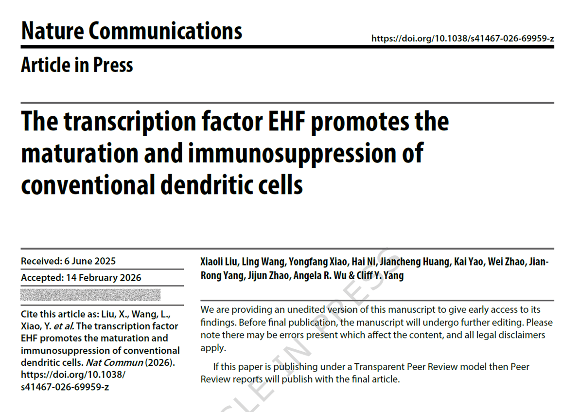
Dendritic cells (DCs) play a critical role in immune regulation, yet the transcriptional mechanisms underlying their immunosuppressive function remain unclear. CCR7⁺ conventional dendritic cells (cDCs) are particularly important in immune tolerance. This study identified ETS homologous factor (EHF) as a key transcription factor regulating cDC maturation and immunosuppression. Mice lacking EHF showed enhanced resistance to autoimmune diseases, infections, and tumors. EHF-deficient cDCs promoted Th1 and Th17 CD4⁺ T cell responses both in vitro and in vivo. EHF can be activated by TLR7/8/9 and inhibited by TLR3, GM-CSF, and IFN-γ. It directly regulates the transcription of genes such as Ccr7, Cd200, Cd274, Irf4, and Rel, promoting the expression of CCR7, CD200, and PD-L1 while suppressing IRF4. Additionally, EHF is highly enriched in CCR7-high DCs in both humans and mice. This study reveals a conserved transcriptional program for cDC-mediated immunosuppression, offering new therapeutic targets for autoimmune diseases, cancer, and infectious diseases.
AtaGenix provided a monoclonal antibody against EHF for this study. The antibody specifically recognizes the EHF protein and served as a specific probe for CUT&TAG analysis, directly supporting the validation of EHF’s transcriptional regulation of target genes and confirming its direct binding to genes such as Ccr7 and Cd200. Meanwhile, the antibody was used in Western blotting and other experiments to verify EHF expression patterns across different tissues and cells, ensuring the reliability of the findings regarding EHF expression and function.
Title: A cell cycle-dependent transition of acetylation to phosphorylation regulates timely centrosome maturation
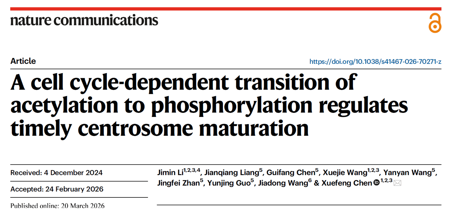
Centrosomes must mature during the G2/M phase to ensure proper bipolar spindle assembly and chromosome segregation. PLK1 is a key kinase in this process, but how the cell cycle regulates its timely recruitment remains unclear. This study found that the ubiquitin E3 ligase RNF40 localizes to centrosomes and is phosphorylated by CDK1 at T529/T557 during G2/M phase. This enables RNF40 to bind PLK1 and promote its recruitment, forming a CDK1-RNF40-PLK1 cascade regulatory mechanism. During interphase, RNF40 is acetylated by PCAF at K517/K562. A transition from acetylation to phosphorylation occurs from late G2 to M phase, which is a prerequisite for cascade activation. Persistent acetylation or phosphorylation defects disrupt PLK1 localization, microtubule nucleation, and bipolar spindle assembly, leading to mitotic catastrophe. Furthermore, RNF40 dysregulation is associated with colorectal cancer. Targeting this cascade may enhance chemotherapy efficacy. This study uncovers a novel cell cycle-dependent mechanism regulating centrosome maturation and provides new targets for cancer therapy.
AtaGenix prepared four site-specific rabbit polyclonal antibodies for this research: anti-pT529-RNF40, anti-pT557-RNF40, anti-acK517-RNF40, and anti-acK562-RNF40. These antibodies precisely detect the phosphorylation or acetylation status of specific sites on RNF40, providing direct evidence for validating RNF40 modification patterns. They ensured the reliability of core data regarding modification sites, modification timing, and functional relevance, forming an important foundation for elucidating the CDK1-RNF40-PLK1 cascade and the biological significance of the acetylation-to-phosphorylation transition.
Title: HE4 drives PD-L1 expression in myeloid cells via IFN-γR-JAK-STAT3 signaling to promote tumor immune evasion
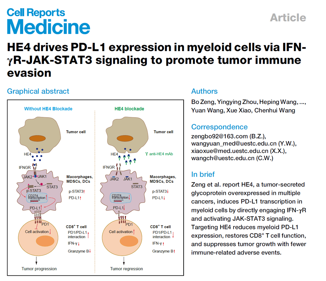
PD-1/PD-L1 immune checkpoint inhibitors are limited by low response rates and immune-related adverse events. The regulatory mechanisms of PD-L1 expression on myeloid cells in the tumor microenvironment remain unclear. This study discovered that tumor-secreted HE4 directly binds to IFN-γR on myeloid cells, activating the JAK-STAT3 signaling pathway, which transcriptionally upregulates PD-L1 expression, suppresses CD8⁺ T cell function, and promotes tumor immune evasion. HE4 competitively binds to IFN-γR with IFN-γ, and high concentrations of HE4 in the tumor microenvironment can antagonize IFN-γ-mediated STAT1 activation. The developed anti-HE4 monoclonal antibody significantly reduced PD-L1 expression on myeloid cells, restored CD8⁺ T cell activity, and inhibited tumor growth in multiple tumor models, with far fewer immune-related adverse events than PD-1 inhibitors. Clinical data showed that high HE4 expression can predict response to PD-1 inhibitors in lung adenocarcinoma patients, making it a potential therapeutic target and predictive biomarker.
AtaGenix prepared rat anti-mouse HE4 monoclonal antibody and mouse anti-human HE4 monoclonal antibody for this study. These antibodies were used to screen clones capable of specifically blocking HE4-induced PD-L1 upregulation, validating HE4’s critical role in PD-L1 regulation. In addition, the antibodies were employed in both in vitro and in vivo tumor inhibition experiments, confirming that HE4 neutralization can reverse the immunosuppressive tumor microenvironment. They also served as key reagents for evaluating the safety and efficacy of HE4 blockade therapy.
Title: Oriented nanobody–field-effect transistor interfaces enable ultrasensitive cancer biomarker detection
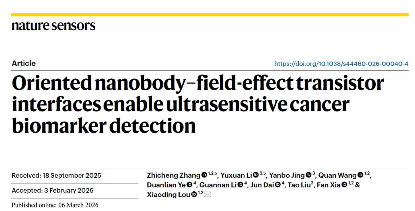
Early screening for endometrial cancer lacks efficient and portable methods. Traditional detection approaches are costly and time-consuming. Although field-effect transistor (FET) biosensors hold great promise, their clinical translation has been hindered by probe instability, non-specific adsorption, and Debye shielding. To address this, the research team developed the SNAP-FET platform, which combines genetic code expansion and click chemistry to achieve site-specific, oriented immobilization of nanobodies on an indium oxide transducer. They also integrated a portable device called ENDOCARE. The platform demonstrated ultra-high sensitivity, specificity, and stability in cell lines, animal models, and clinical samples, showing strong concordance with ELISA results. The ROC curve AUCs for detecting HE4 and CA125 in 18 endometrial cancer patients and 11 healthy individuals were 0.8434 and 0.8131, respectively. This study provides a new strategy for the clinical translation of FET biosensors and lays a technological foundation for point-of-care early tumor diagnosis.
AtaGenix undertook the construction and screening of a CA125 nanobody phage display library for this research. The natural VHH phage display library (1.51 × 10¹⁰ colony-forming units) constructed by AtaGenix served as the core foundation for screening CA125-specific nanobodies. After three rounds of panning and monoclonal screening, a high-affinity CA125-targeting nanobody, NT2, was successfully obtained. This nanobody became a key recognition element for CA125 detection in the SNAP-FET platform, enabling multiplex detection of two major endometrial cancer biomarkers and providing an important probe to validate the universality of the site-specific modification method.
Title: A proliferative subpopulation of coelomocytes in sea cucumber is identified and characterized by single-cell RNA-seq after evisceration
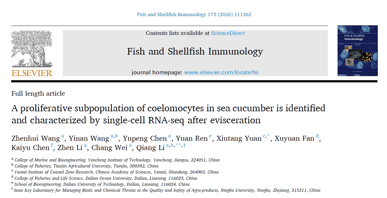
Regeneration research is a cutting-edge global scientific topic. As an echinoderm with remarkable regenerative capacity, the sea cucumber can rapidly regenerate coelomocytes after evisceration, but the underlying recovery mechanisms remain unclear. Using single-cell RNA sequencing, this study analyzed coelomocytes from the coelom and water vascular system of the sea cucumber Apostichopus japonicus, identifying 19 distinct subpopulations. After evisceration, the proportion of subpopulation 10 increased 15-fold in the coelom and 10-fold in the water vascular system. This subpopulation exhibits proliferative characteristics. In the coelom, subpopulation 10 can directly differentiate into subpopulations 0, 4, and 5, while in the water vascular system, it undergoes dedifferentiation before differentiating into subpopulation 9 (immune-related cells). The genes Rplp0 and piwi play key regulatory roles in this process. The study provides a comprehensive molecular resource for classifying sea cucumber coelomocytes and reveals the dynamic changes in their composition and function following evisceration.
AtaGenix provided custom NLRC3 primary antibody preparation services for this experiment. Since NLRC3 serves as a marker gene for subpopulation 9 and lacked suitable sequences for in-house antibody production, AtaGenix synthesized peptides from its highly conserved regions and generated a specific primary antibody. In immunocytochemical staining, this antibody successfully characterized the morphological features of subpopulation 9 cells (diameter 5–6 μm, spherical shape, moderate nucleus-to-cytoplasm ratio, with positive signals appearing as large granules in the cytoplasm). It provided critical experimental evidence for validating the immune function of subpopulation 9 and clarifying its differentiation relationship with subpopulation 10.
Thank you for your attention and support!
AtaGenix will continue to provide high-quality one-stop technical services including custom antibody development and protein expression, empowering more groundbreaking scientific discoveries.

Contact Us

+86-27-65523339

info@atagenix.com

Building C, R & D Building, No. 666, Shendun 4th Road, Donghu New Technology Development Zone, Wuhan